Table of Contents >> Show >> Hide
- What is panniculitis?
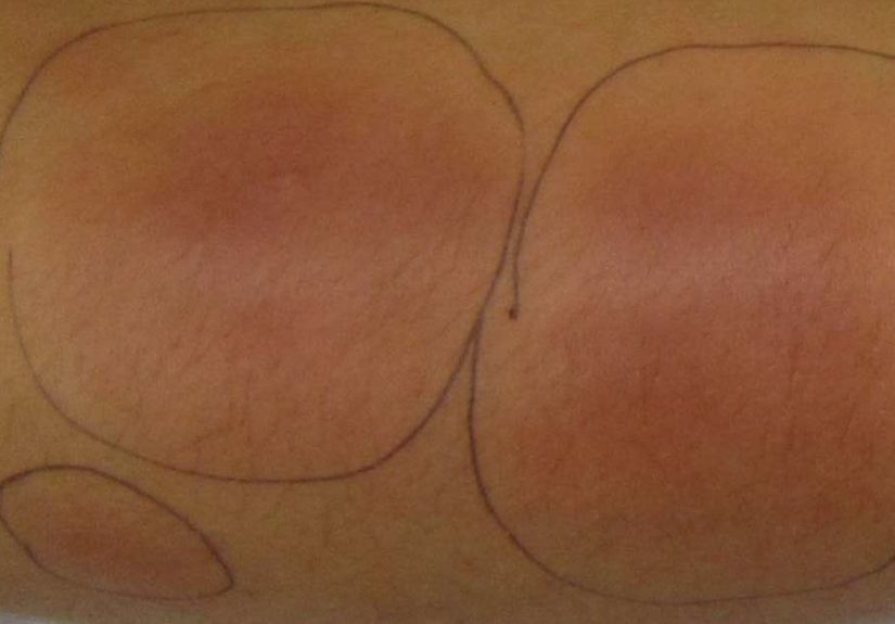
- Symptoms: what panniculitis typically feels and looks like
- What causes panniculitis?
- Types of panniculitis: the big ones you’ll actually hear about
- 1) Erythema nodosum (the “classic” panniculitis)
- 2) Nodular vasculitis / erythema induratum
- 3) Lupus panniculitis (lupus profundus)
- 4) Pancreatic panniculitis
- 5) Alpha-1 antitrypsin deficiency–associated panniculitis
- 6) Cold panniculitis (including “popsicle panniculitis”)
- 7) Infectious panniculitis
- 8) Medication-related or other inflammatory forms
- Diagnosis: how clinicians figure out which type you have
- Treatment: what actually helps (and why it depends on the type)
- Outlook: will it go away?
- Questions to ask at your appointment
- Experiences: What living with panniculitis can feel like
If your skin suddenly starts growing tender “lumps” like it’s auditioning for a bubble-wrap commercial, you’re not aloneand you’re not imagining it.
Panniculitis is a group of conditions that cause inflammation in the fat layer under the skin (subcutaneous fat).
The tricky part: many different problems can trigger it, and several types can look similar at first glance.
This guide breaks down the most common symptoms, the major types of panniculitis, how clinicians figure out which one it is,
and what treatment usually helps. (Spoiler: the best treatment depends on the “why,” not just the “what.”)
Educational note: This article is for general information and isn’t a substitute for medical care. If you have new, painful nodules or fever, get evaluated.
What is panniculitis?
“Panniculitis” is an umbrella term for inflammation in subcutaneous fat. Think of your fat layer as a padded jacket under the skin:
when it gets inflamed, you may feel firm, sore nodules or plaquesoften on the lower legs. Some forms are short-lived and harmless.
Others are clues to infections, autoimmune disease, medication reactions, pancreatic problems, or rare deficiencies.
How panniculitis is classified (why doctors care)
When a clinician suspects panniculitis, a biopsy can help classify the inflammation as:
septal (in the connective tissue “walls” between fat lobules), lobular (inside the fat lobules), and whether there’s
vasculitis (inflamed blood vessels). That pattern narrows the list of causes and guides treatment.
Symptoms: what panniculitis typically feels and looks like
Skin symptoms
- Tender, deep lumps (nodules) or thicker areas (plaques) under the skin
- Red, pink, purple, or bruise-like discoloration over the bumps
- Warmth over the area
- Pain with pressureeven jeans can feel offensive
- Sometimes ulceration (skin breakdown) or oily drainage, depending on the type
Whole-body symptoms
Some types come with systemic symptoms, especially when triggered by infection or autoimmune inflammation:
- Fever, fatigue, “flu-ish” feeling
- Joint pain (ankles and knees are frequent complainers)
- Sore throat or cough (if an infection is involved)
- Digestive symptoms (if inflammatory bowel disease is a trigger)
When to seek urgent care
Panniculitis isn’t usually an emergency, but get prompt medical care if you have:
- Rapidly spreading redness, severe pain, or fever (possible serious infection)
- Open sores, black/purple skin changes, or drainage
- Shortness of breath, chest pain, or leg swelling (other diagnoses can mimic panniculitis)
- Severe abdominal pain, vomiting, or yellowing of the eyes/skin (possible pancreatic or liver-related issues)
What causes panniculitis?
Panniculitis is a “reaction pattern.” The fat layer can become inflamed for lots of reasons, including:
- Infections (bacterial, fungal, tuberculosis and others)
- Inflammatory/autoimmune conditions (like sarcoidosis, lupus, inflammatory bowel disease)
- Medication reactions (some antibiotics, hormonal medications, and others)
- Physical triggers (cold exposure or trauma)
- Pancreatic disease (rare but important because it can show up before belly symptoms)
- Genetic/metabolic issues (such as alpha-1 antitrypsin deficiency in uncommon cases)
Types of panniculitis: the big ones you’ll actually hear about
Below are common and clinically important types, with practical “clues” that often help differentiate them.
(Diagnosis still depends on medical evaluationand sometimes biopsy.)
| Type | Typical clues | Common triggers | Common treatment direction |
|---|---|---|---|
| Erythema nodosum | Tender red nodules, usually on shins; bruise-like fading; often with ankle pain | Strep throat, sarcoidosis, IBD, pregnancy, meds | Supportive care + treat trigger |
| Nodular vasculitis / erythema induratum | Nodules often on calves; may ulcerate; can be linked to vascular inflammation | Historically linked with TB; also other triggers | Treat underlying cause; anti-inflammatory therapy |
| Lupus panniculitis (lupus profundus) | Firm deep nodules that can heal with dents/atrophy; lupus features may be present | Cutaneous or systemic lupus | Often antimalarials + anti-inflammatory meds |
| Pancreatic panniculitis | Nodules on legs; can ulcerate; may precede pancreatic symptoms | Pancreatitis or pancreatic tumor (rare) | Fix the pancreatic problem |
| Alpha-1 antitrypsin deficiency–associated panniculitis | Painful nodules/plaques that can ulcerate and leak oily material | Alpha-1 antitrypsin deficiency (rare) | Specialist-directed therapy + treat deficiency |
| Cold/traumatic panniculitis | Localized inflammation after cold exposure or injury; often self-limited | Ice packs, cold objects; blunt trauma | Avoid trigger + supportive care |
1) Erythema nodosum (the “classic” panniculitis)
Erythema nodosum is the most commonly discussed form of panniculitis. It classically appears as
tender red nodules on the front of the shins, and they often fade through bruise-like colors as they resolve.
People may also notice ankle swelling or joint aches.
A key point: erythema nodosum is often a signnot the main event.
It can be triggered by streptococcal throat infection, sarcoidosis, inflammatory bowel disease,
certain medications (including some antibiotics or hormonal therapies), and pregnancy. Sometimes, no cause is found.
Real-life example: A person gets a sore throat, feels better, and two weeks later develops painful shin nodules and ankle achesclassic timing for a post-infectious trigger.
2) Nodular vasculitis / erythema induratum
This is often described as a more lobular panniculitis pattern and may include inflammation of blood vessels.
Lesions can be more common on the calves and may ulcerate in some cases.
Because triggers vary, evaluation often includes screening for infections (including TB risk when appropriate) and other inflammatory causes.
3) Lupus panniculitis (lupus profundus)
Lupus panniculitis involves inflammation of subcutaneous fat related to lupus.
People can develop firm, deep nodulesoften on the arms, shoulders, buttocks, face, or trunk.
Over time, it can heal with indentations (fat atrophy), which is one reason early recognition matters.
Treatment frequently focuses on controlling lupus-related inflammation. Clinicians often consider
antimalarial therapy (like hydroxychloroquine) and anti-inflammatory medications, with the exact plan tailored to disease severity and overall lupus activity.
4) Pancreatic panniculitis
This rare type is important because skin findings can appear before obvious pancreatic symptoms.
It’s associated with pancreatic inflammation (pancreatitis) or pancreatic cancer in some cases.
Nodules typically show up on the legs and may ulcerate.
The “headline” treatment is to address the underlying pancreatic disease. When the pancreatic issue improves,
the panniculitis often improves as wellanother reason doctors don’t treat this like “just a rash.”
5) Alpha-1 antitrypsin deficiency–associated panniculitis
Alpha-1 antitrypsin (AAT) deficiency is more widely known for lung and liver effects, but a rare skin manifestation is panniculitis.
Lesions can be painful, recurrent, and in some cases ulcerate and leak oily material.
Because this is uncommon and can be stubborn, management is typically specialist-directed and may include therapies that address the underlying deficiency
alongside anti-inflammatory approaches.
6) Cold panniculitis (including “popsicle panniculitis”)
Cold can irritate subcutaneous fatespecially in children, whose fat composition and skin physiology make them more susceptible.
“Popsicle panniculitis” is a classic example: a cold object against the cheek can lead to localized redness and firm bumps.
The good news: it’s usually self-limited. The “treatment” is mainly to stop the cold exposure and use comfort measures.
7) Infectious panniculitis
Some panniculitis is directly caused by infection in or near the fat layer.
This requires quick evaluation because the treatment may involve antibiotics or antifungals and sometimes drainage,
depending on severity and the organism.
8) Medication-related or other inflammatory forms
Certain drugs can trigger panniculitis-like reactions, and there are additional inflammatory variants tied to systemic disease.
That’s why a medication list (including supplements) mattersand why clinicians ask questions that seem unrelated, like GI symptoms, cough, or new joint pain.
Diagnosis: how clinicians figure out which type you have
History and exam: the detective work
Diagnosis starts with timing and context: recent sore throat? new medication? pregnancy? chronic cough? diarrhea?
travel? cold exposure? A clinician will look at location (shins vs calves vs trunk), tenderness, warmth, color changes, and whether lesions ulcerate.
Why a biopsy can be the turning point
When the cause isn’t obviousor when lesions are unusual, persistent, ulcerating, or recurrentclinicians may recommend a
deep skin biopsy that includes subcutaneous fat. Pathology can identify septal vs lobular inflammation and look for vasculitis,
fat necrosis, granulomas, or signs of infection.
Common tests (based on the suspected trigger)
- Inflammation markers: ESR/CRP, CBC
- Infection work-up: throat testing for strep when symptoms fit; TB or fungal evaluation when risk factors exist
- Chest imaging: if sarcoidosis is suspected
- GI evaluation: when symptoms suggest Crohn’s/ulcerative colitis
- Pancreatic labs: amylase/lipase when pancreatic panniculitis is a concern
- Autoimmune testing: when lupus or other rheumatic disease features are present
- AAT testing: if lesions ulcerate with oily drainage or if there’s personal/family history suggesting deficiency
Treatment: what actually helps (and why it depends on the type)
Here’s the most important principle: panniculitis treatment is cause-driven.
Symptom relief matters, but long-term improvement usually comes from treating the trigger.
Step 1: Calm symptoms while you investigate
- Rest and leg elevation (especially with lower-leg involvement)
- Compression stockings if swelling is present and your clinician approves
- NSAIDs for pain and inflammation (when safe for you)
- Warm or cool comfort measures depending on what feels better (avoid ice if cold-triggered)
Step 2: Treat the underlying cause
- Infections: treat the organism (antibiotics/antifungals as directed)
- Medication triggers: stop/replace the offending drug under medical guidance
- Sarcoidosis or IBD: manage the systemic condition (often improves skin findings)
- Lupus panniculitis: control lupus-related inflammation (often antimalarials and other anti-inflammatory therapies)
- Pancreatic panniculitis: address pancreatitis or pancreatic malignancy promptly
- AAT deficiency panniculitis: specialist evaluation for deficiency-directed therapy and inflammation control
Medications you might hear about (not a DIY menu)
Depending on the type and severity, clinicians may consider:
- Corticosteroids (used selectivelyoften when infection has been ruled out and symptoms are significant)
- Colchicine in certain inflammatory patterns
- Potassium iodide in some cases of erythema nodosum under clinician supervision
- Dapsone or other immune-modulating therapies for selected diagnoses
- Hydroxychloroquine and related therapies for lupus-associated disease
These options have real side effects and interactions, so they should be guided by a clinician who knows your diagnosis and health history.
What not to do
- Don’t “power through” severe leg pain with intense exerciserest often helps early erythema nodosum flare-ups.
- Don’t apply prolonged ice packs to suspicious nodules; cold can worsen or even cause cold panniculitis.
- Don’t ignore systemic symptoms (fever, weight loss, persistent cough, abdominal pain). Panniculitis can be a clue to something bigger.
Outlook: will it go away?
Many casesespecially erythema nodosum and cold/trauma-related panniculitisimprove over weeks with supportive care and trigger management.
Recurrence risk depends on whether the underlying cause persists (for example, uncontrolled inflammatory bowel disease or ongoing medication exposure).
Some forms can leave temporary discoloration. A few (notably lupus panniculitis) may lead to lasting contour changes if inflammation damages fat,
which is why evaluation and appropriate treatment matter.
Questions to ask at your appointment
- Which type of panniculitis do you suspect, and why?
- Do I need a deep skin biopsy (and if so, where should it be taken)?
- What underlying causes should we rule out in my case (infection, sarcoidosis, IBD, lupus, pancreatic disease)?
- Which pain/inflammation treatments are safest with my medical history?
- What signs mean I should contact you urgently?
- If this is likely to recur, what prevention steps make sense for me?
Experiences: What living with panniculitis can feel like
Panniculitis has a way of being both dramatic and confusing: the pain can be very real, while the outside appearance may look like “just a bruise.”
Many people describe the first week as a weird mismatch“It doesn’t look that bad, but walking feels like I’m stepping on Lego bricks.”
The tenderness is often deep, and the nodules can feel like small stones under the skin. Clothes that normally don’t registertight socks, denim,
even a bedsheet brushing the shinsuddenly become part of the problem.
Another common experience is the “diagnosis detour.” Because multiple conditions can mimic each other (cellulitis, bruising, blood clots, vasculitis),
people sometimes bounce between urgent care, primary care, and dermatology before getting clarity. Many patients say that the moment a clinician
mentions a deep biopsy is when things start to make sense. It’s not that anyone is being dramatic; it’s that panniculitis lives in the fat layer,
and shallow samples can miss the key pattern. When pathology finally labels something like “septal panniculitis” or “lobular panniculitis with vasculitis,”
the chaos turns into a plan.
People with erythema nodosum often share a similar timeline story: a sore throat, a respiratory infection, a new medication, or a stressful
immune flarethen, a week or two later, the shins revolt. One person might notice ankle swelling first and think they “overdid it” at the gym,
only to find tender nodules the next morning. Another might be managing ulcerative colitis and recognize the skin flare as a sign their gut inflammation
is waking up again. In these cases, the emotional experience can be surprisingly validating: once the underlying trigger is treated or controlled,
the nodules usually calm down, and it feels like the body is finally speaking the same language across organs.
For those with autoimmune-related panniculitis (like lupus panniculitis), the experience is often more on-and-off. People describe it as a
“slow burn” that can flare with stress, sun exposure, or medication changes. Because lupus panniculitis can heal with dents in the skin, some patients
report feeling anxious about visible changesespecially when nodules appear on the arms, face, or torso. Practical coping tips that come up repeatedly
include photographing new lesions (so you can show changes over time), keeping a symptom timeline, and asking clinicians to coordinate care between
dermatology and rheumatology so treatment targets both skin comfort and long-term prevention.
Parents dealing with cold panniculitis (like “popsicle panniculitis”) often describe pure confusionfollowed by relief. A red, firm cheek patch
can look alarming, but once a clinician explains the cold trigger and that it typically resolves, the whole household exhales. The “lesson learned”
is usually practical rather than scary: limit prolonged cold contact, avoid direct ice pack pressure on bare skin, and watch for improvement over weeks.
Across types, the most consistent “real life” theme is this: panniculitis is often a clue. People feel betterphysically and mentallywhen the visit
focuses not only on pain control, but on the bigger question: “What is my body reacting to?” When that question is answered, treatment stops being
guesswork and starts being targeted.