Table of Contents >> Show >> Hide
- Quick reality check: What does “movable” actually mean?
- Common causes of a movable breast lump
- 1) Fibroadenoma (the classic “moves easily” lump)
- 2) Breast cyst (fluid-filled and sometimes dramatic)
- 3) Fibrocystic breast changes (AKA “my boobs have opinions”)
- 4) Fat necrosis (a lump after injury or surgery)
- 5) Infection or inflammation (mastitis, abscess, plugged ducts)
- 6) Less common (but real): Phyllodes tumor and other growths
- 7) Breast cancer (sometimes a lump is the first clue)
- When to worry (and when to book the appointment today)
- Diagnosis: What to expect at the doctor’s office
- Treatment: What happens after you get an answer
- What you can do right now (without spiraling)
- FAQ
- Conclusion
- Experience Corner: What It’s Like to Find a Movable Lump (and Not Lose Your Mind)
You’re in the shower. Your hand does a casual sweep. And thenplot twistyou feel a lump. It moves a little under your fingers, like it’s trying to dodge responsibility. Your brain immediately opens 37 tabs at once: Is this cancer? Is this normal? Is this… a rogue pea?
Take a breath. A movable lump in the breast is very often caused by benign (non-cancerous) conditions. But “often” isn’t the same as “always,” and the only way to know what you’re dealing with is a proper evaluation. This guide breaks down the most common causes, what doctors look for, which tests you might need, and the treatment options without doom-scrolling or medical jargon that sounds like it was invented to win Scrabble.
Quick reality check: What does “movable” actually mean?
When people say a breast lump is “movable,” they usually mean it shifts slightly under the skin when pressed. Many benign lumpsespecially fibroadenomascan feel smooth, rubbery, and mobile. Cysts can also feel like they “roll” a bit, depending on size and location.
Here’s the important part: mobility alone does not diagnose anything. Some cancers can feel movable early on, and some benign lumps can feel firm or less mobile. The goal isn’t to become your own radiologistit’s to notice changes and get the right workup.
Common causes of a movable breast lump
A “breast lump that moves” can come from several conditions. Age, hormone cycles, pregnancy/breastfeeding, recent injuries or procedures, and family history can all shape what’s most likely.
1) Fibroadenoma (the classic “moves easily” lump)
A fibroadenoma is a solid, benign breast tumor and one of the most common reasons a young person finds a distinct lump. It’s often described as firm, smooth, rubbery, round, painless, and mobile. Many fibroadenomas show up in the teens through 30s, but they can occur at other ages too.
What tends to happen next: your clinician confirms the lump on exam and usually orders imaging (often ultrasound). If it looks typical, you may simply monitor it over time. If it’s growing, causing discomfort, or looks unusual, a biopsy or removal may be recommended.
2) Breast cyst (fluid-filled and sometimes dramatic)
Cysts are fluid-filled sacs inside the breast. They can feel soft or firm, tender or painless, and sometimes appear fast like they were waiting for your busy week to strike. Cysts can fluctuate with the menstrual cycle and may be more common in people in their late 30s to 50s.
Ultrasound is especially helpful because it can usually tell whether a lump is fluid-filled (more consistent with a cyst) or solid (which could be a fibroadenoma or something else). Painful cysts can sometimes be drained with a needle.
3) Fibrocystic breast changes (AKA “my boobs have opinions”)
Fibrocystic changes are common and related to hormone fluctuations. People often describe generalized lumpiness, tenderness, or “ropey” textureespecially before a period. Lumps may come and go or feel different month to month.
The tricky part is that new, localized lumps still deserve evaluation. “Common” doesn’t mean “ignore it forever.”
4) Fat necrosis (a lump after injury or surgery)
Fat necrosis can occur after breast trauma (even something you barely remember), surgery, radiation, or injections. It’s benign, but it can form a lump that sometimes feels firm and can mimic other conditions on exam. Imaging helps sort this out.
5) Infection or inflammation (mastitis, abscess, plugged ducts)
If a lump comes with redness, warmth, swelling, fever, or feeling sick, infection jumps higher on the list especially during breastfeeding. Mastitis can cause a tender area or lump, and untreated infection can sometimes progress to an abscess (a pocket of pus) that may need antibiotics and drainage.
Translation: if you feel awful and your breast looks angry, don’t “wait and see” for a week. Get seen promptly.
6) Less common (but real): Phyllodes tumor and other growths
Phyllodes tumors are rare and usually start in connective tissue. Many are benign, but some can be borderline or malignant. They can grow quickly, so a rapidly enlarging lump needs prompt evaluation.
7) Breast cancer (sometimes a lump is the first clue)
Breast cancer can present as a new lump or thickening. While people often picture a hard, fixed, irregular mass, real life is messier: cancers can sometimes feel softer or even movable. That’s why clinicians focus on the whole pictureexam, imaging, and (when needed) biopsy.
When to worry (and when to book the appointment today)
Any new breast lump deserves medical attention, but these features raise urgency:
- New lump that persists beyond one menstrual cycle
- Rapid growth over weeks to months
- Skin changes: dimpling, puckering, thickening, redness that doesn’t improve
- Nipple changes: new inversion, persistent discharge (especially bloody)
- Enlarged lymph nodes in the armpit or above the collarbone
- Systemic symptoms: fever, chills, feeling ill (think infection)
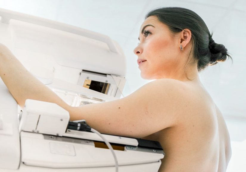
Diagnosis: What to expect at the doctor’s office
Most evaluations follow a simple logic: history → exam → imaging → biopsy (if needed). The goal is to match what the clinician feels with what imaging showsthis “concordance” matters.
Step 1: History and clinical breast exam
Expect questions like: When did you notice it? Does it change with your period? Any pain, nipple discharge, skin changes, pregnancy/breastfeeding, recent injury or surgery, medications (including hormone therapy), and family history. Then they’ll examine both breasts and nearby lymph nodes.
Step 2: Imaging (chosen largely by age and situation)
Imaging isn’t one-size-fits-all. Clinicians often use:
- Ultrasound to evaluate whether a lump is fluid-filled or solid (and it’s commonly used in younger patients)
- Diagnostic mammography (often for age 40+ or when indicated) to assess the area and look for other findings
- Tomosynthesis (3D mammography) in many centers as part of diagnostic workups
Sometimes imaging reports include a BI-RADS category (a standardized risk/next-step system). “Probably benign” findings may be followed with short-interval imaging rather than immediate biopsy, depending on the case.
Step 3: Biopsy (when imaging or exam can’t confidently call it benign)
If the lump is suspicious, growing, or unclear, your clinician may recommend sampling tissue. Common approaches include:
- Fine-needle aspiration (FNA): thin needle, sometimes used for cysts or certain evaluations
- Core needle biopsy: larger needle takes small tissue cores (often image-guided)
- Excisional biopsy: surgical removal, usually reserved for select cases
Many biopsies are guided by ultrasound to target the exact spot. It sounds intimidating, but for most people, it’s an outpatient procedure with local numbingmore “annoying dentist visit” than “movie-scene drama.”
Treatment: What happens after you get an answer
If it’s a fibroadenoma
Many fibroadenomas can be safely monitored with repeat imaging, especially if they’re small and stable. Removal may be considered if a lump is large, painful, growing, cosmetically bothersome, or causing significant anxiety. (Yes, peace of mind counts in real life.)
If it’s a cyst
Simple cysts may need no treatment. If a cyst is painful or large, aspiration (draining with a needle) can relieve symptoms. Complex cysts or uncertain findings may require closer follow-up or biopsy, depending on the imaging features.
If it’s fibrocystic change
Management is often symptom-focused: supportive bras, over-the-counter pain relief if appropriate, and tracking patterns with your cycle. Your clinician may recommend tailored strategies if symptoms are severe or persistent.
If it’s infection (mastitis/abscess)
Treatment depends on severity and whether you’re breastfeeding. It may include supportive measures, antibiotics, andif an abscess formsdrainage. Don’t try to “tough it out” if symptoms worsen or you develop fever.
If it’s phyllodes tumor
Phyllodes tumors are typically treated with surgical removal because they can recur, and some forms can be malignant. Follow-up imaging may be recommended after treatment.
If it’s cancer
Treatment is highly individualized and may include surgery (lumpectomy or mastectomy), radiation, chemotherapy, hormonal therapy, targeted therapy, and/or immunotherapy depending on the cancer type and stage. The key takeaway: earlier evaluation generally expands options and improves outcomes.
What you can do right now (without spiraling)
- Don’t poke it 400 times a day. (You’ll just bruise yourself and stress-test your nervous system.)
- Note the details: location, size (roughly), tenderness, mobility, skin/nipple changes, and timing with your cycle.
- Schedule an exam with a primary care clinician, OB-GYN, or breast specialist.
- Go sooner if there’s fever, redness, rapid growth, nipple discharge, or skin dimpling.
FAQ
Does a movable lump mean it’s not cancer?
Not necessarily. Many benign lumps move easily, but some cancers can also feel mobileespecially early. That’s why imaging (and sometimes biopsy) matters.
Should I wait until after my period to see if it goes away?
If you suspect hormonal changes and you’re not having red-flag symptoms, it’s reasonable to observe briefly. But a new lump still warrants evaluationespecially if it persists beyond one cycle.
What test is best for a palpable lump?
It depends on age, risk factors, and exam findings. Ultrasound is commonly used for many palpable lumps, while diagnostic mammography is often part of evaluationparticularly for people 40 and older.
Conclusion
A movable lump in the breast is often caused by benign conditions like fibroadenomas or cysts, and sometimes it’s related to normal hormonal shifts. But your fingertips can’t issue a final verdictand they shouldn’t have to. A timely medical evaluation can clarify what it is, relieve anxiety, and get you appropriate treatment (or reassurance) faster.
If you found a new lump, your next best step is simple: book the appointment. Your future self will thank youand your search history can finally chill.
Experience Corner: What It’s Like to Find a Movable Lump (and Not Lose Your Mind)
Let’s talk about the part no one puts on the brochure: the moment you notice a movable breast lump, your brain becomes a full-time screenwriter. The plot is always the sameeverything is ominous, the music swells, and suddenly you’re convinced you can feel your pulse in your eyebrows. Here are a few real-world style experiences (the kind clinicians hear every day) that can make the journey feel less lonely.
Experience #1: “It feels like a little marble, and it scoots away when I touch it.”
This is one of the most common descriptions of a fibroadenoma. People often say it’s painless, smooth, and oddly “polite” like it moves just enough to remind you it exists but not enough to be caught in the act. The stress comes from uncertainty: you don’t know if you should ignore it, Google it, or build a tiny shrine of worry around it. In many cases, an ultrasound brings fast clarity: “Yes, this looks benign,” followed by a plan to monitor it. The emotional whiplashfrom panic to reliefcan be intense. Tip from the trenches: write down your questions before the appointment. Anxiety loves to delete your memory the second a clinician says, “Any questions?”
Experience #2: “It showed up overnight and now it hurts.”
Cysts and hormonally related changes can feel like they arrive with dramatic flair. You might notice tenderness, swelling, or a lump that seems to change with your cycle. People often feel frustrated because it’s not just scaryit’s uncomfortable. When a clinician explains that some lumps are fluid-filled and not dangerous, it can feel like getting your brain back. If a cyst is painful, draining it can provide surprisingly quick relieflike popping a balloon you didn’t realize you were holding in your chest. The takeaway: pain doesn’t automatically mean “bad,” but it does mean you deserve evaluation and symptom relief.
Experience #3: “I’m breastfeeding and now there’s a tender lump with redness.”
Breastfeeding adds its own chaos: plugged ducts, mastitis, and (sometimes) abscesses. People often describe feeling sick, feverish, and guiltylike they somehow caused it. They didn’t. Infections happen, and early care helps. What surprises many is how quickly symptoms can escalateand how quickly treatment can help. Emotional tip: ask for clear “if/then” guidance. For example: “If my fever isn’t better in 24 hours, what should I do?” Having a plan reduces late-night spirals.
Experience #4: “My lump moves, but I’m still terrified.”
This is more common than you think. Even when clinicians say a lump looks benign, your mind may keep whispering, “But what if…?” That’s not irrational; it’s your protective instincts working overtime. Many people feel better after a follow-up scan confirms stability, or after a biopsy definitively labels the tissue. If you’re losing sleep, tell your clinician. Anxiety is a symptom too, and your care plan can include reassurance strategiesnot just medical ones.
The big lesson from most experiences: the worst part is often the in-between timeafter you’ve found the lump, but before you have answers. Your best tools in that window are practical: schedule the visit, stop repeatedly checking the lump, and lean on credible information rather than internet rabbit holes. You’re not “overreacting.” You’re responding to uncertainty. The goal is to replace uncertainty with a diagnosis and a plan.