Table of Contents >> Show >> Hide
- What Is Tumoral Thrombosis (Tumor Thrombus)?
- Why It Matters: Tumor Thrombus vs Regular Blood Clot
- Symptoms: What Tumoral Thrombosis Can Feel Like
- Causes and Risk Factors
- Diagnosis: How Doctors Confirm It (and Tell What Type It Is)
- Treatment: What Options Look Like (and Why They Differ)
- Complications and Prognosis
- Living With Tumoral Thrombosis: Practical Questions to Ask Your Care Team
- Real-World Experiences (Patient, Caregiver, and Clinician Perspectives)
- Experience #1: “They Found It on a Scan I Almost Didn’t Want” (Kidney Cancer Scenario)
- Experience #2: “It Wasn’t Just the TumorIt Was the Blood Flow” (Liver Cancer Scenario)
- Experience #3: The Caregiver View“I Needed a Checklist More Than a Pep Talk”
- Experience #4: The Clinician Perspective“The Scan Tells a Story, But It’s Not a Novel”
- Conclusion: The Takeaway
“Tumoral thrombosis” sounds like a villain from a medical drama, but it’s a real (and seriously important) condition. It usually refers to a tumor thrombus: cancer cells growing into a blood vessel and forming a mass inside the vein. Sometimes people also use the phrase to mean blood clots in people with cancer (cancer-associated thrombosis). Either way, the headline is the same: something is blocking blood flowand the next steps depend on what that “something” actually is.
This guide breaks down symptoms, causes, how doctors tell a tumor thrombus from a regular clot, and what treatment can look likewith clear examples (and a tiny bit of humor, because medicine is stressful enough already).
What Is Tumoral Thrombosis (Tumor Thrombus)?
A tumor thrombus is tumor tissue inside a blood vessel. It’s not just “sticky blood.” It’s an extension of the cancer itselfoften growing from the main tumor directly into a nearby vein.
The classic examples:
- Kidney cancer (renal cell carcinoma) growing into the renal vein and sometimes up the inferior vena cava (IVC) (the body’s main “return-to-heart” vein).
- Liver cancer (hepatocellular carcinoma) growing into the portal vein (the vein bringing blood to the liver).
- Less commonly: certain lung cancers, pancreatic neuroendocrine tumors, sarcomas, and some childhood tumors.
Meanwhile, a “bland” thrombus is the usual blood clot made of platelets and fibrinwhat most people picture when they hear “clot.” Cancer can raise the risk of bland clots too, so some patients have both tumor thrombus and bland clot at the same time. That’s where diagnosis gets spicy (in a “please let this be straightforward” kind of way).
Why It Matters: Tumor Thrombus vs Regular Blood Clot
The reason doctors care so much about the difference is simple: a tumor thrombus often needs cancer-directed treatment (surgery, radiation, targeted therapy, immunotherapy), while a bland clot usually needs anticoagulation (“blood thinners”). Using the wrong playbook can mean wasted timeor unnecessary bleeding risk.
Quick Comparison
| Feature | Tumor Thrombus | Bland (Typical) Thrombus |
|---|---|---|
| What it is | Tumor cells growing inside a vessel | Blood clot (platelets/fibrin) |
| Often connected to a tumor? | Yesfrequently contiguous with the mass | Not necessarily |
| Imaging behavior | May enhance with contrast; may expand the vein; may show internal flow | Typically non-enhancing; may shrink with time/therapy |
| Primary treatment | Cancer treatment (sometimes surgery/thrombectomy) | Anticoagulation and risk-factor management |
| Response to blood thinners | Often persists if it’s truly tumor tissue | Often improves/resolves (not always, but often) |
Symptoms: What Tumoral Thrombosis Can Feel Like
Symptoms depend on which vessel is involved and whether there’s also a bland clot. Many tumor thrombi are found incidentally on imaging done for cancer stagingmeaning the patient didn’t feel a “clot symptom” at all.
Common Clot-Style Symptoms (DVT/PE)
- Leg or arm swelling (often one-sided), pain/tenderness, warmth, redness or discoloration
- Shortness of breath, chest pain (especially with deep breaths), fast heartbeat, coughing up blood (possible pulmonary embolism)
Symptoms by Location (Tumor Thrombus “Traffic Jams”)
- Renal vein/IVC (common in kidney cancer): leg swelling, visible abdominal veins, scrotal swelling, fatigue, kidney-area pain, blood in urine (sometimes from the tumor itself).
- Portal vein (common in liver cancer): abdominal swelling (ascites), worsening liver function, enlarged spleen, and complications of portal hypertension such as variceal bleeding.
- Hepatic veins (Budd-Chiari–type picture): abdominal pain, ascites, enlarged liver, liver dysfunction.
- Superior vena cava (rare, often lung/mediastinal tumors): facial/neck swelling, shortness of breath, prominent chest veins.
When to Treat This Like an Emergency
Seek urgent care immediately for sudden shortness of breath, chest pain, fainting, coughing blood, or rapidly worsening swelling. Those can signal a pulmonary embolism or other urgent complications.
Causes and Risk Factors
Tumoral thrombosis is usually about how the cancer growsbut cancer also makes the blood more likely to clot in general. Think of it as a double feature: the tumor can invade the vessel, and the body can be in a hypercoagulable state.
Why a Tumor Thrombus Happens
- Direct invasion: Some cancers grow into nearby veins (kidney tumors are famous for this).
- Venous drainage pathways: Tumor cells can extend along veins that drain the tumor.
- Local vessel effects: Tumors compress vessels, slowing flow and encouraging clot formation around them.
Why Cancer Raises “Regular Clot” Risk (Cancer-Associated Thrombosis)
- Tumor-driven clotting signals (procoagulant activity and inflammation)
- Immobility, hospitalizations, dehydration
- Major surgery (especially cancer surgery)
- Central venous catheters and ports
- Certain therapies (some chemotherapy regimens and other systemic treatments can increase clot risk)
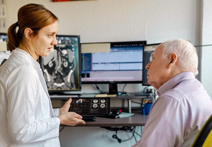
Diagnosis: How Doctors Confirm It (and Tell What Type It Is)
Diagnosing tumoral thrombosis is part detective work, part high-tech photography. Clinicians combine symptoms, cancer history, physical exam, andmost importantlyimaging.
History and Exam
- New swelling, pain, shortness of breath, or abdominal distension
- Known cancer type and stage (some cancers are more “vessel-invading” than others)
- Bleeding risk factors (critical for treatment planning)
Blood Tests (Helpful, but Not the Star of the Show)
Tests like D-dimer can be hard to interpret in cancer because levels may be elevated for many reasons. Bloodwork is still useful to assess anemia, platelets, kidney/liver function, and baseline clotting statusespecially before treatment.
Imaging: The Main Event
- Doppler ultrasound: great for limb clots and sometimes abdominal vessels; can show flow patterns.
- CT with contrast: common for staging and can reveal thrombus, vessel expansion, and relation to the tumor.
- MRI with contrast: especially helpful for defining the extent of tumor thrombus and characterizing tissue.
- PET/CT: can help in selected situations if the tumor type is metabolically active.
- Echocardiography: sometimes used if tumor thrombus is suspected to extend near the heart.
Imaging Clues That Suggest Tumor Thrombus
- Contiguity: the thrombus appears connected to the main tumor mass
- Enhancement: it “lights up” with contrast like tumor tissue
- Vessel expansion: the vein may look enlarged by the mass
- Internal vascularity/flow: features suggesting living tissue rather than inert clot
- Persistence despite anticoagulation: on follow-up imaging, a true tumor thrombus often doesn’t shrink like a bland clot might
Sometimes it’s still ambiguous on a single scan. In those cases, doctors may rely on serial imaging (watching how it changes over time) and the broader clinical picture.
Treatment: What Options Look Like (and Why They Differ)
Treatment depends on three big questions: (1) Is it tumor thrombus, bland clot, or both? (2) Where is it? (3) What’s the overall cancer plan and bleeding risk?
1) Cancer-Directed Treatment (Core for Tumor Thrombus)
Because a tumor thrombus is tumor tissue, the main approach is treating the cancer. Options vary by cancer type and stage, but may include:
- Surgery to remove the primary tumor and the tumor thrombus (often used in kidney cancer when feasible)
- Systemic therapy (targeted therapy, immunotherapy, chemotherapydepending on the cancer)
- Radiation therapy in selected scenarios (sometimes used to control local disease or reduce symptoms)
- Locoregional liver therapies for hepatocellular carcinoma with portal vein involvement in appropriate candidates (approach depends on liver function, extent of disease, and guideline strategy)
Example: Kidney Cancer with IVC Tumor Thrombus
In renal cell carcinoma, tumor thrombus can extend into the renal vein and up the inferior vena cava. When surgery is appropriate, the treatment may involve radical nephrectomy plus tumor thrombectomy. These can be technically complex operations that sometimes require a multidisciplinary team (for example, urologic oncology with vascular or cardiac surgery involvement), depending on how far the thrombus extends.
Example: Liver Cancer with Portal Vein Tumor Thrombosis
Portal vein involvement often signals more advanced disease and can limit options like transplant in many cases. Management frequently leans on systemic therapy and, in selected patients, liver-directed treatments aimed at controlling tumor burden and maintaining liver function. The plan is highly individualized because the liver’s reserve (how well it still works) matters as much as the tumor does.
2) Anticoagulation (“Blood Thinners”): Sometimes, But Not Always
Here’s the non-glamorous truth: anticoagulation for tumor thrombus is a gray zone. A bland clot is made to be dissolved and stabilized by anticoagulation. A tumor thrombus is living tissueblood thinners don’t “melt” tumor.
Clinicians may consider anticoagulation when:
- There’s evidence of mixed thrombus (tumor thrombus plus bland clot)
- The patient has a separate DVT/PE elsewhere
- There’s high concern for clot propagation and the bleeding risk is acceptable
They may avoid or use extra caution when:
- Bleeding risk is high (for example, certain GI cancers, low platelets, or advanced liver disease)
- The thrombus appears to be purely tumoral and anticoagulation would add risk without clear benefit
3) Interventional and Supportive Options
- IVC filters: sometimes considered when anticoagulation is impossible and PE risk is high from a bland clot component (not a default choice, and they can complicate future procedures).
- Symptom management: pain control, compression strategies (when appropriate), mobility support, and monitoring.
- Managing portal hypertension complications: if portal vein involvement leads to varices/ascites, treatment often targets those complications directly.
What Follow-Up Often Includes
- Repeat imaging to monitor thrombus size/behavior
- Ongoing cancer treatment response assessment
- Bleeding/clotting monitoring if anticoagulated
- Coordination across oncology, surgery, hematology, radiology, and sometimes hepatology
Complications and Prognosis
A tumor thrombus can raise the stakes because it may:
- Indicate a more locally advanced cancer (affecting staging and treatment planning)
- Cause obstruction-related symptoms (swelling, organ congestion, portal hypertension)
- Coexist with bland clots, increasing the risk of DVT/PE
Prognosis depends heavily on the cancer type, extent of thrombus, whether disease has spread elsewhere, and whether the tumor (and thrombus) can be effectively treated or removed. Importantly, some scenariossuch as surgically removable kidney cancer with venous tumor thrombuscan still have meaningful outcomes when treated appropriately.
Living With Tumoral Thrombosis: Practical Questions to Ask Your Care Team
If you or a loved one is dealing with tumoral thrombosis, these questions can help clarify the plan:
- Is the thrombus tumoral, bland, or mixed? What features support that?
- What’s the goal of treatment? Cure, control, symptom relief, or preventing complications?
- Do I need anticoagulation? If yes, which medication and for how longand what bleeding signs should I watch for?
- What symptoms should send me to the ER?
- How will we monitor it? What imaging, and when?
- Which specialists should be involved? (Oncology, hematology, surgery, hepatology, etc.)
Also: bring a full medication list. Blood thinners, supplements, and cancer therapies can interact in surprisingly dramatic wayslike a group text that should’ve stayed one-on-one.
Real-World Experiences (Patient, Caregiver, and Clinician Perspectives)
The medical facts matter, but so does the human part: tumoral thrombosis can feel confusing because it sits at the intersection of “cancer problem” and “clot problem.” Many people describe the early days as a blur of new terms, urgent scans, and questions that start with, “Wait… is this the kind of clot blood thinners fix?”
Experience #1: “They Found It on a Scan I Almost Didn’t Want” (Kidney Cancer Scenario)
A common story in kidney cancer is that the tumor thrombus shows up during staging imaging. The patient may have had vague fatigue, back discomfort, or blood in the urinesymptoms that are easy to brush off until a CT scan tells the plot twist. The phrase “tumor thrombus” can sound terrifying, especially when someone explains it can extend toward the heart. People often recall two emotions arriving at once: fear, and relief that there’s a concrete plan.
When surgery is recommended, patients frequently talk about how surreal it feels to learn that removing a kidney can be paired with removing a “plug of tumor” from a major vein. Many say the most helpful moment was a specialist calmly drawing a diagram: kidney → renal vein → IVC → heart, with a simple line showing where the thrombus ends. That visual turns a scary concept into something you can discuss, question, and understand.
Experience #2: “It Wasn’t Just the TumorIt Was the Blood Flow” (Liver Cancer Scenario)
For people with liver cancer and portal vein tumor thrombosis, the experience can be different: symptoms may be driven less by a limb swelling “clot” and more by liver-related changesabdominal bloating, fluid buildup, appetite loss, and fatigue that feels like walking through wet cement. Patients often describe learning a new vocabulary: “portal vein,” “portal hypertension,” “varices,” and “liver reserve.”
Many find it emotionally tough that certain treatments may be limited by liver function. The upside is that care teams often focus on two goals at once: controlling the tumor and protecting quality of life. People frequently say they felt more in control once they understood the “why” behind each test: imaging to track tumor burden, labs to track liver function, and careful discussion of bleeding risk if anticoagulation is on the table.
Experience #3: The Caregiver View“I Needed a Checklist More Than a Pep Talk”
Caregivers often become the unofficial project managers of modern medicine. A helpful pattern many adopt is a short running list: What kind of thrombus is it? What are the red-flag symptoms? Who do we call after hours? When is the next scan? In stressful moments, the brain is not a filing cabinetit’s a browser with 47 tabs open and one of them is playing music. Writing down medication names, doses, and the team’s “if X happens, do Y” instructions can reduce panic and prevent mistakes.
Experience #4: The Clinician Perspective“The Scan Tells a Story, But It’s Not a Novel”
Radiologists and clinicians often describe tumor thrombus diagnosis as pattern recognition plus context. Enhancement, vessel expansion, and continuity with the tumor can strongly suggest tumoral thrombosis, but borderline cases happen. Providers may emphasize follow-up imaging because time reveals behavior: a bland clot might shrink with anticoagulation; tumor tissue often doesn’t.
Many clinicians also stress shared decision-making: when bleeding risk is high, “just anticoagulate” isn’t a reflexit’s a careful conversation. Patients who do best, clinicians say, are the ones who feel comfortable asking direct questions and repeating them until the plan makes sense. If you ever worry you’re “being annoying,” remember: clarity is not annoying; it’s safety.
Conclusion: The Takeaway
Tumoral thrombosis is not just “a clot.” Often, it’s tumor growth inside a vesselespecially in cancers like renal cell carcinoma and hepatocellular carcinoma. Symptoms range from none at all (found on scans) to significant swelling, pain, breathing trouble, or abdominal complications depending on the vessel involved. Diagnosis relies heavily on contrast imaging and clinical context, and treatment usually centers on cancer-directed therapysometimes with surgerywhile anticoagulation is reserved for selected situations (particularly when bland clot is also present).
If you remember one thing, make it this: the right treatment depends on the right label. “Tumor thrombus” and “blood clot” can look similar but behave differently, and your care team’s job is to figure out which one you’re dealing withthen act fast and wisely.