Table of Contents >> Show >> Hide
- What Is a Childhood Glioma?
- Types of Childhood Glioma
- Causes and Risk Factors: What We Know (and What We Don’t)
- Symptoms of Childhood Glioma
- How Childhood Glioma Is Diagnosed
- Treatment Options: The Toolbox (and How Doctors Choose)
- Prognosis and Life After Treatment
- Questions to Ask Your Child’s Care Team
- Real-Life Experiences: What Families Often Describe (and Why It Matters)
- Final Thoughts
Let’s start with an unfair truth: the word “tumor” can turn a normal Tuesday into a
“what even is time?” kind of day. If you’re here because a doctor mentioned glioma (or because you
Googled “morning headaches + vomiting” at 2 a.m.), take a breath. Childhood gliomas are a broad
group of brain and spinal cord tumors, and outcomes range from “highly treatable” to “we need a
very serious game plan.” The key is knowing what you’re dealing withand why your child’s care
team keeps saying, “It depends on the location.”
This guide breaks down types, causes and risk factors,
symptoms, diagnosis, and treatment optionsplus what
families often experience in real life (the parts nobody puts on a clinic brochure).
Medical note: This article is for education, not a diagnosis. If you’re worried about urgent symptoms (like a first seizure, severe lethargy, or rapidly worsening weakness), seek emergency care.
What Is a Childhood Glioma?
A glioma is a tumor that starts in the brain or spinal cord from glial cellsthe nervous system’s
support crew. Think of neurons as the “main characters” sending signals, and glial cells as the team handling
insulation, nutrition, cleanup, and structure so the show can go on.
In kids, gliomas are often discussed by grade (how aggressive the cells look and behave) and by
where the tumor is. That location piece matters because the brain is not a single empty roomit’s a packed
city. A small tumor in a sensitive spot can cause major symptoms, while a larger one elsewhere might stay quiet longer.
Low-grade vs. high-grade (in plain English)
- Low-grade gliomas tend to grow more slowly and are often very treatablesometimes even curable, especially if surgery can remove the tumor fully.
- High-grade gliomas grow faster, can spread into nearby brain tissue, and usually need a combination of treatments.
Types of Childhood Glioma
“Glioma” is an umbrella term. Under it are several tumor types, and the names can sound like they were invented
during a spelling bee held in a wind tunnel. Here are the big categories families hear most often.
1) Pediatric Low-Grade Glioma (pLGG)
These are the most common gliomas in children and usually grow slowly. Many start in the cerebellum
(the back-of-the-brain “coordination headquarters”). Treatment can range from watchful waiting to surgery, chemo,
radiation, or newer targeted medicines.
- Pilocytic astrocytoma (very common low-grade type)
- Diffuse low-grade glioma / diffuse astrocytoma
- Optic pathway glioma (affects vision pathways; sometimes linked with NF1)
- Subependymal giant cell astrocytoma (SEGA) (often linked with tuberous sclerosis)
2) Pediatric High-Grade Glioma (pHGG)
High-grade gliomas in children are less common than low-grade tumors, but they’re more aggressive and often require
a multi-step plan (surgery when possible, plus radiation and chemotherapy, sometimes with targeted or immune-based
approaches in trials). The exact subtype and molecular features matter a lot.
- Anaplastic astrocytoma (higher grade)
- Glioblastoma (very high-grade)
- Infant-type hemispheric glioma (a distinct entity in very young children; often driven by specific gene fusions)
3) Diffuse Midline Glioma (DMG), H3 K27-altered (often called DIPG when in the pons)
This is a high-grade glioma that grows in midline brain structures such as the thalamus or brainstem. When it’s in the
pons, it’s commonly known as diffuse intrinsic pontine glioma (DIPG). These tumors are challenging because they sit in
areas that control essential functions like breathing, swallowing, and movement.
4) Spinal Cord Gliomas
Gliomas can also form in the spinal cord. Symptoms may include back pain, weakness, sensory changes, or problems with walking.
Treatment depends on the tumor type, location, and whether surgery can be done safely.
A quick comparison table
| Type | Typical Growth | Common Locations | Often-Discussed First Steps |
|---|---|---|---|
| Low-grade glioma (e.g., pilocytic) | Slow | Cerebellum, optic pathway, cerebral hemispheres | Observation vs. surgery; then chemo/targeted therapy if needed |
| High-grade glioma | Fast | Cerebral hemispheres, sometimes brainstem | Surgery when feasible; radiation + chemo; consider trials |
| Diffuse midline glioma (H3 K27-altered) | Fast, infiltrative | Brainstem/pons, thalamus, spinal cord | Biopsy may guide molecular therapy; radiation is common |
Causes and Risk Factors: What We Know (and What We Don’t)
Parents often ask, “What caused this?” and the honest answer is: most childhood gliomas happen without a clear cause.
Gliomas develop when changes occur in how glial cells grow and divide, but the exact trigger for those changes is often unknown.
What medicine can identify are a few risk factors that raise the odds.
Inherited genetic conditions
Some children have a higher risk because of inherited conditions. Two that come up frequently are:
- Neurofibromatosis type 1 (NF1) (associated with optic pathway gliomas and other low-grade tumors)
- Tuberous sclerosis (associated with certain low-grade tumors, including SEGAs)
Prior radiation exposure (in specific medical contexts)
Exposure to radiation therapy to the brain in the past can raise the risk of developing brain tumors later. This is not the same as normal day-to-day
“screen time radiation” (which is not considered a proven cause). It’s about medical radiation used to treat a condition.
What about “everyday” causes?
It’s human nature to look for a single villainphones, microwaves, a bonk on the head, that one time your child ate a whole bag of
neon-blue candy. But for most children, there’s no evidence of a specific, preventable cause. If guilt is trying to move into your brain rent-free,
it doesn’t deserve the key.
Symptoms of Childhood Glioma
Symptoms depend on location, size, and growth rateplus your child’s age and development.
Some tumors cause symptoms quickly; others are found incidentally on a scan done for another reason.
General symptoms (often related to pressure in the skull)
- Morning headaches or headaches that improve after vomiting
- Nausea and vomiting (especially persistent or unexplained)
- Vision changes (blurred vision, crossed eyes)
- Balance problems or trouble walking
- Behavior or personality changes or unusual sleepiness
- Seizures
Symptoms by location (the “map matters” part)
- Cerebellum: clumsiness, wobbliness, frequent falls, trouble with coordination
- Optic pathway/hypothalamus: vision loss, abnormal eye movements, hormonal or growth changes
- Frontal/temporal lobes: personality changes, headaches, seizures, language or memory changes
- Brainstem (including DIPG/DMG): double vision, facial weakness, trouble swallowing, changes in walking or speech
- Spinal cord: back pain, weakness, numbness/tingling, changes in bladder/bowel function
Symptoms in babies and toddlers
Younger children can’t always describe headaches or vision issues. Signs may include increasing head size, persistent vomiting,
unusual sleepiness, developmental regression, or a new preference for “not walking today” when they used to zoom around like a tiny CEO.
Important perspective: most of these symptoms can also be caused by common illnesses. The concern is pattern and persistencesymptoms that
worsen over time, are worst in the morning, disrupt sleep, or come with new neurologic changes.
How Childhood Glioma Is Diagnosed
Diagnosis typically starts with a careful history and neurologic examchecking vision, hearing, strength, balance, coordination, reflexes,
and sometimes school performance or behavior changes (yes, “my kid is suddenly struggling in math” can be a real clue).
Imaging: MRI is the workhorse
For suspected brain tumors, MRI is the main imaging test. It helps show tumor location, size, and relationship to surrounding structures.
A follow-up MRI after surgery often checks how much tumor remains.
Biopsy and tumor testing
Some tumors can be removed surgically, allowing the team to examine tumor tissue. In other casesespecially when the tumor sits in a very sensitive location
doctors may do a biopsy to confirm the diagnosis and run molecular testing. This testing looks for genetic changes that can guide treatment,
including targeted therapies.
Special case: NF1-associated optic pathway glioma
In some children with NF1, an optic pathway glioma can be diagnosed based on clinical findings and imaging, and a biopsy may not be needed.
Treatment decisions often focus on preserving vision rather than “treating the scan.”
Treatment Options: The Toolbox (and How Doctors Choose)
Treatment is tailored to the child and the tumor. The same word “glioma” can mean very different plans, from “watch and wait” to a multi-modality approach.
Pediatric brain tumor care is usually handled by a team that may include pediatric neuro-oncology, neurosurgery, radiation oncology, neurology, endocrinology,
rehabilitation, neuropsychology, and school support.
1) Observation (watchful waiting)
Some low-grade gliomasespecially those found incidentally or those associated with NF1may be monitored with regular MRIs and exams.
This isn’t “doing nothing.” It’s “choosing the safest next step while keeping a close eye on the plot.”
2) Surgery
When safe and feasible, surgery aims to remove as much tumor as possible. For certain low-grade tumors, a complete resection can be curative.
If a tumor is in a high-risk location, the surgeon may remove only part of it to reduce symptoms and protect function.
3) Chemotherapy
Chemotherapy is commonly used for tumors that can’t be fully removed or that regrow. In younger children, chemo may also be used to
delay radiation, because the developing brain is more sensitive to radiation’s long-term effects.
4) Radiation therapy (including proton therapy)
Radiation can be effective for controlling gliomas, especially higher-grade tumors and some recurrent or progressive low-grade tumors.
Techniques such as conformal approaches and proton beam therapy may help reduce radiation exposure to nearby healthy tissue.
The timing and type of radiation are individualized.
5) Targeted therapy (precision medicine)
In recent years, targeted therapies have changed the landscape for some pediatric gliomasespecially low-grade tumors driven by changes in pathways like MAPK.
Depending on the tumor’s molecular profile, doctors may consider options such as:
- MEK inhibitors (used in certain low-grade gliomas, including some NF1-associated or BRAF fusion-driven tumors)
- BRAF-targeted therapies (for tumors with a BRAF V600 mutation or certain BRAF fusions)
- mTOR inhibitors (notably for SEGAs in tuberous sclerosis)
Targeted therapy isn’t “magic,” but it can offer tumor shrinkage or control with a different side-effect profile than traditional chemo or radiation.
Long-term monitoring remains important because kids are still growing, and so is the science.
6) Diffuse midline glioma / DIPG: what’s different
For diffuse midline glioma (including DIPG in the pons), surgical removal isn’t typically possible because the tumor infiltrates vital brain structures.
A biopsy may still be done to confirm diagnosis and identify molecular features. Radiation therapy is commonly used to slow progression and
ease symptoms, and many families explore clinical trials.
7) Clinical trials
Clinical trials are a major part of pediatric neuro-oncology, especially for tumors where standard options are limited.
Large cooperative groups and research networks help test new strategiessometimes new drug combinations, sometimes precision medicine approaches,
sometimes immunotherapies. Your care team can help you evaluate whether a trial fits your child’s specific diagnosis and goals.
Supportive care matters (a lot)
Gliomas and treatments can cause symptoms like headaches, nausea, fatigue, weakness, vision changes, hormonal issues, and learning challenges.
Supportive care can include steroids to reduce swelling, anti-seizure medications when needed, physical/occupational therapy, speech therapy,
neuropsychology support, and help with school accommodations.
Prognosis and Life After Treatment
Prognosis depends on tumor type, location, how much can be removed, and molecular features. Many children with low-grade glioma do very well,
though some tumors behave like long-term conditions that require periodic treatment and monitoring. High-grade gliomas and diffuse midline gliomas are
generally more difficult, and outcomes can vary widely based on subtype and treatment response.
Follow-up often includes regular MRIs, neurologic exams, and screening for late effectsespecially if radiation or certain chemotherapies were used.
Survivorship care may also address learning, attention, mood, endocrine health, vision, hearing, and physical functioning. The goal isn’t just survival;
it’s helping a child live a full, thriving life in the body and brain they have today.
Questions to Ask Your Child’s Care Team
- What exact type of glioma is this, and what grade is it?
- Where is it located, and how does that affect symptoms and treatment options?
- Is surgery possible? If not, whyand what are the alternatives?
- Was molecular testing done? Are there targetable genetic changes?
- What’s the goal right now: cure, long-term control, symptom relief, or all of the above?
- What side effects should we watch for (short-term and long-term)?
- How often will MRIs happen, and what counts as “growth that needs action”?
- Are there clinical trials appropriate for this diagnosis?
- What support is available for school, therapy, mental health, and siblings?
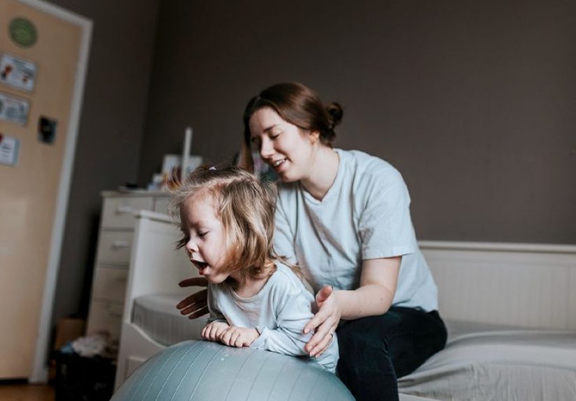
Real-Life Experiences: What Families Often Describe (and Why It Matters)
The medical facts are important, but families also live in the space between appointments: the ordinary days that suddenly feel unfamiliar. Many parents describe
a “slow realization” periodweeks or months where something seems off, but not in a dramatic, movie-scene way. It might be a headache that keeps returning, a kid
who suddenly hates bright light, or a once-steady runner who starts clipping door frames like the hallway moved. Often, it’s the patternnot one symptom
that finally triggers an urgent call.
A common theme is the symptom calendar. Families jot down: “Headache before school,” “vomited after waking,” “fell twice at soccer,” “teacher said handwriting changed.”
This isn’t overreacting; it’s good detective work. Patterns can help clinicians decide when imaging is needed. And just as importantly, the act of tracking can make you feel a tiny bit
less powerlesslike you’re holding a flashlight in a very dark room.
Then there’s MRI day, which deserves its own emotional category. Some kids do great; others hate the noise and the stillness. Parents often talk about the surreal
contrast: the child in superhero pajamas, the high-tech scanner, and the grown-ups trying to act calm while their brains are sprinting. If your child needs sedation, families describe
a mix of relief (“they’ll be comfortable”) and nerves (“I hate this part”). It can help to ask the team exactly what to expecthow long, what recovery looks like, and who you can call
if post-sedation nausea or grogginess lingers.
If the diagnosis is a low-grade glioma, many families experience a mental whiplash: “It’s a tumor” colliding with “It might be watched for now.” Observation can feel emotionally harder
than treatment because there’s no visible “doing.” Parents often say the first follow-up scan is the toughest. Over time, many develop a rhythm: scan dates on the calendar, a plan for
distraction, and a trusted point person on the care team for questions. It becomes less like living in constant emergency mode and more like managing a chronic condition with check-ins.
For families facing high-grade glioma or diffuse midline glioma, the experiences can be heavier: rapid symptom changes, intense treatment schedules, and hard conversations about goals of care.
Many caregivers talk about learning a new languageradiation fractions, steroids, clinical trial criteriaand also learning how to protect moments of childhood in the middle of medical life.
That can look like decorating the hospital room, celebrating “small wins” (a good appetite day counts!), or insisting on normal rituals like bedtime stories, even if the setting is different.
Across all types, families often emphasize the value of support that isn’t strictly medical: social workers who help with school plans, child life specialists who make procedures
less scary, therapists who give kids a place to be angry without worrying about hurting their parents, and sibling support that prevents brothers and sisters from becoming invisible background characters.
If you’re reading this as a caregiver, you don’t have to be “strong” 24/7 to be effective. You just have to keep showing upand accept help when it’s offered.
One last shared experience: the “new normal” isn’t a single destination. It’s a series of adjustmentssometimes two steps forward, one step back. Many families say the best coping skill is
building a short list of trusted questions, then re-asking them whenever the plan changes. Medicine evolves, tumors change, kids grow, and what was true last year might not be true next scan.
It’s okay to need things explained more than once. Confusion is not a character flaw; it’s a normal response to a high-stakes situation.