Table of Contents >> Show >> Hide
- First Things First: What “Recovery” Really Means
- Common Treatments and Why They Change Your Timeline
- The Detached Retina Recovery Timeline at a Glance
- What “Normal” Recovery Symptoms Can Look Like
- Positioning: Why It Matters (and How to Survive It)
- Restrictions: The Big “Don’ts” (and the Reason Behind Them)
- Medication and Home Care: Your “Boring but Powerful” Routine
- Follow-Up Visits: The Quiet Hero of Good Outcomes
- Red Flags: Call Your Eye Doctor ASAP If You Notice These
- Vision Recovery: Why It Can Take Weeks to Months
- Outlook and Prognosis: What Actually Influences the Long-Term Result
- FAQs People Google at 2 a.m.
- Real-World Recovery Experiences (Extra 500+ Words)
- Experience #1: “The Bubble Is a Floating Mood Ring”
- Experience #2: “Face-Down Positioning Makes Time Weird”
- Experience #3: “My Vision Improves… Then Freaks Me Out… Then Improves Again”
- Experience #4: “The Other Eye Starts Doing All the Work”
- Experience #5: “The Anxiety Is Real (and So Is the Relief)”
- Experience #6: “Later Surprise: Cataract Haze After Vitrectomy”
- Wrap-Up: The Big Takeaways
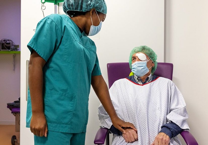
A detached retina is one of those medical situations where your eye basically waves a tiny red flag and says,
“Hey, I’m not being dramatic… but I am an emergency.” The good news: modern retinal surgery is highly effective.
The slightly annoying news: recovery has its own personalitypart science, part patience, and part learning to sleep
in a position that makes you feel like a rotisserie chicken.
This guide walks you through what recovery typically looks like after retinal detachment repair, what the timeline can
feel like day-by-day and month-by-month, and what influences long-term outlookwithout turning your brain into a medical
textbook. (Your retina has been through enough.)
First Things First: What “Recovery” Really Means
Recovery after a detached retina is not just “the eye feels better.” It’s a layered process:
the retina is reattached and sealed, inflammation settles, the eye pressure stabilizes, the brain adapts to changes in
vision, and your visual system slowly recalibrates. Some people notice improvement within weeks; others need months.
And in certain casesespecially if the macula (your sharp central-vision area) detachedvision may improve but not fully
return to what it was before.
Translation: success often means your retina is back where it belongs and your vision is protected from getting worse.
It does not always mean instant HD vision the next morning. If your expectations are realistic, your stress level will be
lowerand your recovery will feel more manageable.
Common Treatments and Why They Change Your Timeline
Your surgeon picks a repair method based on the type of detachment, tear location(s), lens status, scarring risk, and
other eye factors. These are the big three you’ll hear about:
1) Pneumatic Retinopexy (Gas Bubble + Positioning)
Often done in-office for select detachments. A gas bubble is placed inside the eye to gently press the retina back into
position, while laser or freezing treatment (cryopexy) seals the tear. Recovery usually hinges on strict head positioning
for a period of time (sometimes many hours per day) so the bubble sits exactly where it needs to. The bubble gradually
shrinks and disappears over days to weeks.
2) Scleral Buckle (The “Support Belt” Around the Eye)
A scleral buckle is a band placed around the outside of the eye to relieve pulling on the retina and support reattachment.
It can be used alone or combined with other techniques. Early recovery can involve soreness and swelling, and vision may
shift because the buckle subtly changes eye shape. Many people return to routine activities within weeks, but vision can
keep improving over months.
3) Vitrectomy (Removing the Vitreous Gel)
A vitrectomy removes the vitreous gel that may be tugging on the retina. The surgeon may place a gas bubble or silicone oil
to hold the retina in place while it heals. If you have a gas bubble, positioning instructions and travel restrictions can
be a major part of recovery. Vitrectomy recovery can be smoother than it sounds, but the vision timeline can be slowerplus
cataract progression is common after vitrectomy in many adults, especially over age 50.
The Detached Retina Recovery Timeline at a Glance
Every eye is unique, but most recoveries follow a recognizable arc. Here’s a practical, “what this might feel like”
timeline. (Your doctor’s instructions always win if they differ.)
| Time Period | What You May Notice | What You Should Usually Do |
|---|---|---|
| Day 0–1 |
Blurry vision, scratchy sensation, watery eye, mild to moderate discomfort. If a gas bubble was used, your vision may look like you’re peering through a moving “dark circle” or a weird horizon line. |
Rest. Use the eye shield if instructed. Start drops exactly as prescribed. Avoid rubbing the eye. Follow any positioning instructions like they’re a VIP pass. |
| Days 2–7 |
Swelling and redness often peak early then slowly improve. Vision may still be very blurryespecially with a bubble. Light sensitivity and tearing are common. |
Keep up with drops, hygiene, and positioning. Avoid heavy lifting/straining. Attend your first follow-up(s). Ask about safe sleep positions and showering. |
| Weeks 2–4 |
Discomfort usually decreases significantly. If you had a gas bubble, it may be noticeably smaller. You may start to see more “around” it. Vision can fluctuate day-to-day. |
Gradually resume light activities if cleared. Protect the eye. Continue drops. Avoid high-impact exercise. Don’t fly or change altitude with a gas bubble. |
| Months 1–3 |
Many people see meaningful improvement, but reading fine print can still be challenging. If the macula detached, central vision may improve slowly and remain distorted for a while. |
Keep follow-ups. Discuss new glasses timing (often delayed until vision stabilizes). Report new flashes/floaters promptly. |
| Months 3–12 |
Vision often continues improving in small increments. Some eyes stabilize by 3–6 months; others take longer. If cataract develops after vitrectomy, vision can get hazier over time until treated. |
Continue routine monitoring. Address cataract or pressure issues if they arise. Protect both eyes and manage risk factors. |
What “Normal” Recovery Symptoms Can Look Like
After retinal detachment repair, it’s common to have redness, irritation, mild pain, tearing, and blurry vision. If you had
a gas bubble, your vision may resemble looking through a lava lamp that refuses to be aesthetic. You might also notice:
- Wavy or distorted vision (especially if the macula was involved)
- Light sensitivity and glare
- Uneven focus between eyes that makes depth perception feel “off”
- Floaters that slowly settle (but any sudden increase should be reported)
- Dryness or gritty sensation from surface healing and drops
One underrated part of recovery is your brain adapting to the new visual input. Even when the retina is physically healed,
the “software update” in your visual processing can take time.
Positioning: Why It Matters (and How to Survive It)
If your surgeon used a gas bubble, positioning isn’t a quirky suggestionit’s physics. The bubble floats, and your head
position determines where it presses. That pressure helps keep the retina in place while the laser/cryo seal strengthens.
Positioning can be required for hours a day and sometimes for up to about a week (occasionally longer depending on the case).
Practical Tips That Make Positioning Less Miserable
- Rent or borrow positioning equipment (face-down chairs and cushions can be game-changers).
- Set timers for breaks approved by your surgeonmental fatigue is real.
- Podcasts and audiobooks become your new best friends.
- Prep your home so essentials are within reach. You don’t want to go treasure-hunting while face-down.
- Ask your doctor about safe sleep positions. “Wing it” is not the vibe here.
Restrictions: The Big “Don’ts” (and the Reason Behind Them)
No Flying (and Watch Altitude) If You Have a Gas Bubble
Air pressure changes can cause a gas bubble to expand and dangerously raise eye pressure. That’s why air travel, mountain
trips, and sometimes even high-elevation driving are restricted until the bubble is fully gone. If your life involves travel,
ask your surgeon what’s safe based on the exact gas used and your healing progress.
Avoid Heavy Lifting and Straining Early On
Many surgeons recommend avoiding heavy lifting and intense exercise while the eye heals, especially during the first few
weeks. The goal is to reduce pressure spikes and inflammation that can interfere with healing. Your doctor will tell you
when it’s safe to resume workoutsand which kinds are okay first.
Tell Every Medical Provider If You Have an Eye Gas Bubble
This one is crucial: certain anesthesia gases (notably nitrous oxide) can expand an intraocular gas bubble and cause a rapid,
dangerous rise in eye pressure. If you need dental work, emergency surgery, or anything involving sedation, you must tell
the provider that you have (or recently had) a retinal surgery gas bubble.
Medication and Home Care: Your “Boring but Powerful” Routine
Most people go home with prescription eye drops (often antibiotic and anti-inflammatory, sometimes pressure-lowering drops).
These drops aren’t optional; they’re how you keep inflammation down and reduce infection risk. Typical home-care basics include:
- Use drops exactly as directed (set alarmsfuture you will be grateful).
- Wash hands before touching the area around your eye.
- Avoid rubbing or pressing on the eye.
- Wear the shield at night if instructed (sleepy you is not a careful you).
- Ask when it’s safe to shower and how to keep water out of the eye early on.
Follow-Up Visits: The Quiet Hero of Good Outcomes
Follow-ups are how your surgeon checks retinal position, eye pressure, inflammation, and the status of any bubble or oil.
Many people are seen within the first day or two after surgery, then again over the next weeks and months depending on the case.
These appointments matter because some complications are most treatable when caught early.
Red Flags: Call Your Eye Doctor ASAP If You Notice These
Some symptoms are “normal recovery.” Others are your eye sending an urgent email with the subject line: PLEASE READ NOW.
Seek urgent care if you have:
- Sudden increase in floaters or flashes
- A new shadow/curtain in your vision
- Significant worsening of vision after it had been improving
- Severe pain, severe headache, nausea/vomiting (possible high eye pressure)
- Increasing redness with thick discharge or fever (possible infection)
Vision Recovery: Why It Can Take Weeks to Months
The retina is delicate nerve tissue. Even after it’s reattached, the photoreceptors and retinal layers may need time to
recover function. If the macula stayed attached (a “macula-on” detachment), outcomes are often better, and the goal is to
repair quickly to preserve sharp vision. If the macula detached (“macula-off”), central vision can still improve after repair,
but it may be slower and less completethink “gradual upgrades,” not an instant reboot.
Also, your final vision depends on more than anatomy. Healing quality, scarring, swelling, eye pressure, and whether you need
additional procedures all shape the final picture. It’s common for doctors to wait before finalizing a glasses prescription,
because early refractions can become inaccurate as the eye stabilizes.
Outlook and Prognosis: What Actually Influences the Long-Term Result
Retinal detachment surgery is generally very successful at reattaching the retina, but sometimes more than one procedure is
needed. Visual outcome depends heavily on:
- Macula status: whether the macula was detached and for how long
- Speed of treatment: earlier repair is typically associated with better outcomes
- Extent of detachment: larger detachments can be more complex
- Scarring risk (PVR): scar tissue can cause traction and redetachment
- Other eye conditions: high myopia, prior cataract surgery, diabetes, etc.
Many people resume most normal activities after healing, though some need vision correction changes, additional monitoring,
or later treatment for issues like cataract (especially after vitrectomy). Your surgeon’s “outlook” discussion is usually
based on your retina’s condition at surgerynot just the procedure nameso two people can have the same surgery and very
different recovery experiences.
FAQs People Google at 2 a.m.
When can I drive again?
Driving depends on vision clarity, comfort, and whether you still have a gas bubble (which can severely limit vision).
Many people can’t safely drive in the early days. Your surgeon will tell you when you meet legal and practical driving
standards. When in doubt: don’t risk it.
When can I go back to work?
Desk work might be possible within 1–2 weeks for some people, but positioning requirements can make that unrealistic.
Physically demanding jobs may require more time. If your job involves driving, lifting, or high-risk environments, expect
a longer pause and a clearer “return-to-work” plan from your doctor.
Will my vision go back to normal?
Sometimes it gets very closeespecially if the macula stayed attached and the repair was prompt. Other times, vision improves
but remains slightly blurry, distorted, or less sharp. The primary goal is saving vision and preventing further loss.
Real-World Recovery Experiences (Extra 500+ Words)
Below are common experiences people report during detached retina recovery. These aren’t one person’s story or medical advice
think of them as patterns that show up again and again in real life, so you don’t feel blindsided by the emotional rollercoaster
or the “is this normal?” moments.
Experience #1: “The Bubble Is a Floating Mood Ring”
People with a gas bubble often describe seeing a shifting dark circle or a sloshing horizon line that changes when they move
their head. Early on, it can feel like trying to read through a teaspoon of ink. Then the bubble shrinksslowlyand you start
seeing more “above” it. Many patients say this phase is oddly encouraging: each day the bubble line drops a little, and it’s a
visual reminder that healing is happening (even if your patience is not).
Experience #2: “Face-Down Positioning Makes Time Weird”
Positioning can be the hardest partnot because it’s painful, but because it’s relentless. People often underestimate the
mental fatigue: neck stiffness, boredom, and that claustrophobic feeling when your day revolves around gravity. A common tip
is to treat positioning like a job with tools: specialized pillows, rented equipment, audiobooks, and a schedule. Those who
plan ahead tend to feel more in control, which matters a lot when recovery feels slow.
Experience #3: “My Vision Improves… Then Freaks Me Out… Then Improves Again”
Fluctuation is a theme. Many patients report a few good days followed by a day where the eye feels irritated or vision seems
worse, especially after increased activity, poor sleep, or missed lubrication. This doesn’t always mean something is wrong;
inflammation can ebb and flow. The key difference is trend and severity. Gradual overall improvement with mild ups and downs
is common. A sudden new curtain or a dramatic drop in vision? That’s a call-your-doctor moment.
Experience #4: “The Other Eye Starts Doing All the Work”
If one eye is blurry for weeks, your brain leans hard on the better-seeing eye. People often complain of headaches, eye strain,
and fatigue from the constant mismatch. Some find that using larger text, better lighting, and frequent breaks helps. Others
find it emotionally challenging because the world feels slightly “off” in depth and clarity. This is one place where compassion
helps: you’re not being dramaticyour visual system is literally rebalancing.
Experience #5: “The Anxiety Is Real (and So Is the Relief)”
Retinal detachment can be scary, and it’s common to feel anxious during recoveryespecially at night, when every tiny sensation
seems louder. Many people describe a turning point after the first or second follow-up appointment, when they hear the retina is
still attached. That reassurance often drops stress levels dramatically. If anxiety lingers, it can help to write down symptoms
and questions for visits, so your mind isn’t constantly running emergency simulations.
Experience #6: “Later Surprise: Cataract Haze After Vitrectomy”
Some patients feel like they’re doing everything right, vision is improving, and thenmonths laterthings start looking foggy
again. In many adults, this ends up being cataract progression after vitrectomy rather than the retina detaching again. People
often describe it as glare, dull colors, or a smudged-window effect. The reassuring part: cataracts are common and treatable,
and many patients report a big jump in clarity after cataract surgery once the eye is stable and cleared by the retina specialist.
The most consistent “experience” across nearly all recoveries is this: detached retina healing is rarely a straight line. It’s a
series of small winsless redness, a lower bubble line, fewer distortions, more comfortable readingstacked over weeks and months.
If you track progress weekly instead of hourly, it usually feels less maddening.
Wrap-Up: The Big Takeaways
Detached retina recovery is a marathon disguised as a medical appointment. Early care (especially positioning and restrictions)
protects the repair. Weeks 1–4 are about healing and stability. Months 1–3 often bring noticeable improvement. And months 3–12
can deliver gradual gains, with occasional detours like cataract or pressure management.
If you’re in recovery right now, here’s the most practical mantra: follow instructions, don’t improvise travel or exercise,
keep follow-ups, and speak up fast if symptoms suddenly change. Your retina is doing precision workgive it the calm, boring
consistency it needs to stick the landing.