Table of Contents >> Show >> Hide
- What Exactly Is Fibrosarcoma?
- Symptoms: What People Usually Notice First
- Causes and Risk Factors
- How Fibrosarcoma Is Diagnosed
- Treatment Options: What “Plan A” Usually Looks Like
- Risks and Complications: The Stuff Nobody Puts on a Billboard
- Prognosis: What Affects Outlook
- Life After Treatment: Follow-Up, Rehab, and Moving Forward
- Frequently Asked Questions
- Real-World Experiences: What It Feels Like (and What People Wish They’d Known)
- Conclusion
Most lumps are innocent. Some are just your body’s way of saying, “Hey, I made a bump.” But every now and then, a lump shows up with big main-character energyand it deserves a proper medical cameo.
Fibrosarcoma is one of those rare diagnoses that can sound like a spell from a fantasy novel, except it’s very real, very medical, and very important to catch and treat correctly. It’s a type of sarcoma (a cancer of connective tissues) that grows from fibroblastscells that normally help form fibrous tissue like tendons and ligaments.
In this guide, we’ll break down fibrosarcoma symptoms, how doctors diagnose it, what treatment usually looks like, what risks to know about, and what life can feel like on the other side. We’ll keep it accurate, in-depth, and just fun enough that you won’t feel like you’re reading a user manual for anxiety.
Note: This article is for education only and does not replace advice from your medical team.
What Exactly Is Fibrosarcoma?
Fibrosarcoma is a malignant (cancerous) tumor made up of fibroblasts, the cells that produce collagen and support structures in the body. It’s considered a soft tissue sarcoma, although it can also involve areas near or within bone.
Adult-type vs. infantile fibrosarcoma
Doctors commonly describe two broad buckets:
- Infantile (congenital) fibrosarcoma (IFS): Usually appears in babies and very young children. Despite being malignant, it often behaves less aggressively than many adult sarcomas and can respond well to treatment.
- Adult-type fibrosarcoma: A rare tumor seen more often in adults. Treatment typically follows soft tissue sarcoma principles (surgery first when possible, plus radiation and/or chemo depending on risk).
Where it shows up
Fibrosarcoma can develop in many locations, but it’s often found in the deep soft tissues of the extremities (arms/legs) or trunk. In kids, it may appear as a mass in an arm or leg. Wherever it lives, its favorite hobby is expanding into nearby tissueso early evaluation matters.
Symptoms: What People Usually Notice First
The most common early clue is simple: a new lump or a lump that’s getting bigger over time. The tricky part is that sarcoma lumps can be painlessmeaning your body doesn’t always send an “ouch” notification.
Common fibrosarcoma symptoms
- A growing mass (often deep, firm, and gradually enlarging)
- Swelling in the affected area
- Pain or soreness if the tumor presses on nerves, muscles, or organs
- Limited movement if it’s near a joint or within tight tissue compartments
- Limping or trouble using an arm/leg (more common in children)
“Red flag” features of a lump
You don’t need to panic over every bump, but you do want to get checked sooner if a mass is:
- Growing over weeks to months
- Larger than a golf ball (size matters in sarcoma workups)
- Deep (not just under the skin)
- Persistent and not clearly linked to an injury that’s resolving
- Causing symptoms like pain, numbness, weakness, or swelling
Symptoms by location (because the body is not one-size-fits-all)
A fibrosarcoma in your thigh may show up as a firm mass you notice when you sit or exercise. A tumor in the abdomen or retroperitoneum (deep belly space) can be sneakiersometimes causing bloating, discomfort, early fullness, or bowel/urinary issues if it presses on organs.
Causes and Risk Factors
Here’s the honest answer: in most cases, doctors can’t point to a single “cause” of fibrosarcoma. Cancer is usually a mix of genetic changes and bad luck, not a moral failing or a consequence of eating one suspicious gas-station hot dog.
Known or suspected risk factors (mostly shared with other soft tissue sarcomas)
- Inherited cancer syndromes: Certain inherited disorders are associated with increased sarcoma risk. If you have a strong family history of early cancers, genetic counseling may be worth discussing.
- Prior radiation therapy: Radiation used to treat another cancer can increase the risk of developing a sarcoma later, usually years after exposure.
- Environmental exposures (uncertain and often overstated online): Some chemical exposures are linked to certain sarcoma types, but for soft tissue sarcomas overall, evidence varies and is not always definitive.
Bottom line: most people with fibrosarcoma don’t have a clear risk factor. That’s why focusing on early detection (recognizing a growing lump and getting it evaluated) often matters more than hunting for a perfect explanation.
How Fibrosarcoma Is Diagnosed
A key rule: if a mass might be a sarcoma, you want the evaluation done thoughtfully. The biopsy approach and surgical planning can affect outcomes, especially if a tumor is deep or near important structures.
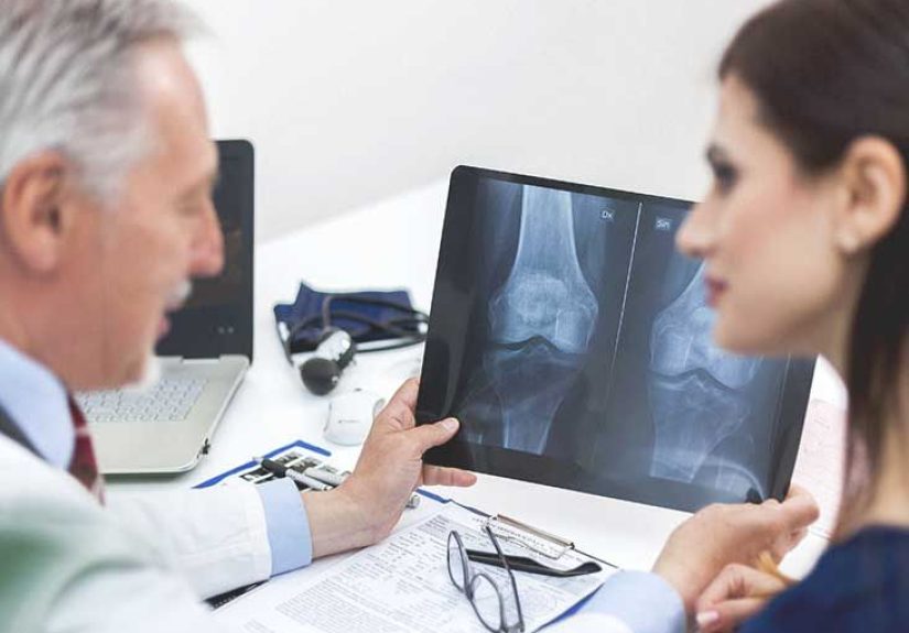
Step 1: Imaging (aka “let’s map this thing”)
For suspected soft tissue sarcoma, imaging commonly includes:
- MRI for masses in arms/legs and soft tissues (great for defining boundaries and relationships)
- CT for certain locations (like abdomen) and to assess for spread
- Chest imaging (often CT) because sarcomas can spread to the lungs
Step 2: Biopsy (the part where the diagnosis becomes real)
A biopsy is essentialimaging alone can’t confirm fibrosarcoma. Many sarcoma workups favor core needle biopsy, which takes small cylinders of tissue for a pathologist to examine. In some situations, a surgical biopsy may be needed.
Step 3: Pathology and (sometimes) molecular testing
Under the microscope, fibrosarcoma is a spindle-cell tumor with specific patterns. But modern sarcoma diagnosis often goes beyond “what it looks like” and includes immunohistochemistry and genetic/molecular testing to classify the tumor accurately. That matters because different sarcoma subtypes can behave differently and respond to different treatments.
Step 4: Staging and grading
Sarcomas are assessed by:
- Grade (how aggressive the cells appear)
- Size and depth
- Location
- Spread to lymph nodes or distant organs (metastasis)
These details guide treatment planning and help estimate recurrence risk.
Treatment Options: What “Plan A” Usually Looks Like
Fibrosarcoma treatment typically follows established soft tissue sarcoma treatment strategies. Translation: the main goal is to remove or control the tumor locally and reduce the chance it comes back or spreads.
Surgery: the cornerstone
Surgery is often the primary treatment when the tumor can be removed safely. Surgeons aim for “clear margins,” meaning they remove the tumor along with a rim of healthy tissue to reduce the chance of leaving cancer cells behind.
For extremity tumors, care teams often pursue limb-sparing surgery when feasible, sometimes combining radiation and/or chemotherapy to preserve function.
Radiation therapy: before or after surgery
Radiation therapy may be used:
- Before surgery (neoadjuvant) to shrink the tumor and improve resectability
- After surgery (adjuvant) to reduce local recurrence risk if margins are close or the tumor is high-grade
Your team will weigh the benefits against side effects (like stiffness, skin changes, swelling, or wound-healing issues), which vary based on dose and location.
Chemotherapy: selective, not automatic
Chemotherapy is not used for every fibrosarcoma. It’s more likely considered when:
- The tumor is high-grade
- There’s metastatic disease (spread)
- The tumor can’t be fully removed at diagnosis
- It’s part of a multimodality plan in certain pediatric cases
Sarcoma chemo regimens vary, and your oncologist tailors decisions to tumor biology, location, and your overall health.
Targeted therapy (especially in infantile fibrosarcoma)
One of the biggest “modern medicine wins” in this space: some infantile fibrosarcomas have an NTRK gene fusion. If testing finds that fusion, doctors may use TRK inhibitors (targeted therapies such as larotrectinib or entrectinib) to shrink tumorssometimes dramaticallymaking surgery easier or less deforming.
This is a perfect example of why molecular testing can matter: it can open a door that didn’t exist a decade ago.
Why sarcoma centers matter
Sarcomas are rare. That means experience is concentrated in specialized centers that use a multidisciplinary team (surgical oncology, orthopedic oncology, radiation oncology, medical oncology, radiology, pathology, rehab). If you’re facing fibrosarcoma, getting a second opinion at a sarcoma-focused center is often a smart movenot because your local team isn’t talented, but because repetition builds expertise, and sarcoma is not the place to crowdsource guesses.
Risks and Complications: The Stuff Nobody Puts on a Billboard
Even with excellent care, fibrosarcoma can come with real risks. Understanding them helps you plan, advocate, and cope.
Local recurrence
Sarcomas can recur in the same area, especially if margins are close, the tumor is high-grade, or it’s in a location where wide surgery is difficult. That’s why surgery planning and (sometimes) radiation matter so much.
Metastasis
Some fibrosarcomas can spread to distant sitescommonly the lungs for many soft tissue sarcomas. Risk depends on grade, size, and biology.
Treatment side effects
- Surgery: scarring, weakness, limited mobility, nerve injury, wound issues
- Radiation: skin irritation, stiffness/fibrosis, swelling/lymphedema, wound-healing concerns
- Chemotherapy: fatigue, nausea, infections (low blood counts), hair loss, neuropathy (depending on drugs)
- Targeted therapy: often better tolerated than classic chemo, but still may cause fatigue, GI symptoms, or lab changes
The sneaky risk: “scanxiety”
Follow-up scans can be emotionally hard. The fear of recurrence is common and valid. Support groups, counseling, and straightforward coping strategies (like scheduling something pleasant after scan day) can make a surprisingly big difference.
Prognosis: What Affects Outlook
Prognosis in fibrosarcoma depends on several factorsnot just the diagnosis itself:
- Tumor grade (higher grade generally carries higher risk)
- Size and depth (larger/deeper tumors can be harder to fully remove)
- Location (some areas allow wider surgery than others)
- Surgical margins (clear margins reduce recurrence risk)
- Metastasis at diagnosis
- Age and overall health
Infantile fibrosarcoma often has a more favorable course than many adult sarcomas, and newer targeted therapies can be game-changing when an NTRK fusion is present. For adults, early evaluation and expert local control (surgery ± radiation) are central to improving outcomes.
Life After Treatment: Follow-Up, Rehab, and Moving Forward
After treatment, most people enter a surveillance phase: regular visits, physical exams, and periodic imaging. The goal is to catch recurrence early and manage long-term effects.
Rehab is not “optional homework”
Physical therapy and occupational therapy can help restore strength, flexibility, and functionespecially after limb surgery or radiation. If you feel like a joint became a rusty door hinge, that’s not you being dramatic. That’s biology.
Practical tips that actually help
- Keep a symptom log (new pain, swelling, or a new lump deserves attention)
- Ask for a survivorship plan (what scans, how often, and what symptoms should trigger a call)
- Protect your mental health like it’s part of treatment (because it is)
- Move your body within your team’s guidanceactivity supports recovery and mood
Frequently Asked Questions
Is fibrosarcoma curable?
Many cases can be treated successfully, especially when the tumor is localized and can be removed with clear margins. “Curable” depends on grade, stage, and locationso it’s a personalized answer, not a one-line fortune cookie.
Does fibrosarcoma spread fast?
It can, but behavior varies. Higher-grade tumors have a greater risk of metastasis. Some low-grade tumors grow more slowly and mainly threaten locally by invading nearby structures.
If it doesn’t hurt, can it still be serious?
Yes. Many soft tissue sarcomas are painless early on. Pain often shows up later, when a tumor presses on nerves or muscles.
Do I need a sarcoma specialist?
If fibrosarcoma or any sarcoma is suspected, a sarcoma-experienced team can help with biopsy planning, pathology accuracy, surgical strategy, and access to the right combinations of therapy.
Real-World Experiences: What It Feels Like (and What People Wish They’d Known)
The stories below are composites drawn from common experiences reported by patients and caregivers in clinical settingsshared here to make the journey feel less abstract and more human. Details are generalized to protect privacy.
Experience #1: “It was just a weird lump… until it wasn’t.”
One adult noticed a firm bump in the thigh that didn’t hurt, didn’t change color, and didn’t do anything dramaticso it got ignored. Months later, it was clearly bigger. The first appointment was reassuring (“Probably benign”), but imaging raised eyebrows. A core needle biopsy confirmed a sarcoma diagnosis, and suddenly the calendar filled up with MRI appointments, consults, and a surgeon who used the phrase “wide margins” like it was a love language.
The biggest surprise wasn’t the surgeryit was how many specialists showed up. Surgical oncology coordinated with radiology and pathology. A physical therapist joined early to plan rehab. The patient later said, “I thought treatment was one doctor and a scalpel. It was a whole orchestra.”
What helped most: writing down questions before every visit, bringing a friend to take notes, and asking the team to explain the plan in plain English: What’s the goal? What are we trying to prevent? What’s the backup plan if margins aren’t clear?
Experience #2: A parent’s shock with infantile fibrosarcoma
In pediatric cases, the emotional whiplash can be intense. A parent may notice a rapidly growing mass on a baby’s arm or leg, assume it’s a cyst, and then hear the word “cancer” in the same week. Many families describe the diagnosis phase as a blur of acronyms, new faces, and a lot of waiting for pathology results.
When molecular testing identifies an NTRK fusion, targeted therapy can become part of the conversation. Some parents describe it as the first moment they felt genuine relief: “There was a specific button the doctors could push.” Tumor shrinkage can make surgery less extensive, which matters a lot for growing bodies.
What helped most: insisting on clear timelines (When will we have results? When do we decide?), asking about function-preserving options, and connecting with a hospital social worker who could translate logisticshousing, work leave, insuranceinto something manageable.
Experience #3: Living with follow-up scans and rebuilding confidence
After treatment, many people expect to feel instantly “back to normal.” Instead, they often discover a new normal: scars, stiffness, occasional pain, and periodic scans that can turn even the calmest person into a professional overthinker.
People commonly describe “scanxiety” as a physical sensationtight chest, racing thoughts, doom-scrolling symptoms at 2 a.m. The most useful coping tools tend to be boring (which is good news, because boring is doable):
- Scheduling scans early in the day to reduce hours of anxious waiting
- Planning a small reward afterward (coffee with a friend, a movie, a nap with no guilt)
- Doing rehab consistently, because regaining strength restores a sense of control
- Joining a support group or seeing a counselor for cancer-related anxiety
A recurring theme: people feel better when they understand the logic of follow-up. Surveillance isn’t “we think something is wrong.” It’s “we’re staying one step ahead.” When teams explain that clearly, follow-up feels less like a threat and more like a safety net.
If you’re in the middle of this: it’s okay to be scared, frustrated, or exhausted. It’s also okay to laugh when something is absurd (like the 14-page packet on how to park). Your job is not to be brave 24/7. Your job is to show up, ask questions, and let a qualified team do their work.
Conclusion
Fibrosarcoma is rare, but the strategy for dealing with it is refreshingly straightforward: take a growing lump seriously, get proper imaging, confirm diagnosis with biopsy, and treat with an expert planoften centered on surgery, sometimes supported by radiation and/or chemotherapy, and increasingly guided by molecular testing (especially in pediatric cases).
If there’s one takeaway: don’t self-diagnose a persistent, growing mass as “just a weird muscle knot.” Let professionals evaluate it. Early, accurate diagnosis and specialized care can make a big difference in treatment choices, function, and peace of mind.